Search This Blog

Showing posts with label Cardiology. Show all posts

Saturday, April 30, 2016

Easy way to learn ECG. Step by Step in a very easy way

Source: Making sense of the ECG


Always start by confirming the name and date of birth of the patient to confirm the ECG belongs to the right person. Also, confirm the date and time the ECG was performed.
Step 1 – Heart rate
Heart rate can be calculated simply with the following method:
  • Work out the number of small squares in one R-R interval
  • Then divide 300 by this number and you have your answer
e.g. If there are 4 squares in an R-R interval 300/4 = 75 beats per minute
.
If the rhythm is irregular:
  • Count the number of complexes on the rhythm strip (each rhythm strip is 10 seconds long)
  • Multiply the number of complexes by 6 (giving you the average number of complexes in 1 minute)
What’s a normal heart rate?
  • Normal = 60 – 100 bpm
  • Tachycardia > 100 bpm
  • Bradycardia < 60 bpm
Hint: If there are obviously P waves present, check the ventricular rate and the atrial rate. The rates will be the same if there is 1:1 AV conduction.
Step 2 – Heart rhythm
The heart rhythm can be regular or irregular.
Irregular rhythms are regularly irregular (i.e. a recurrent pattern of irregularity) or irregularly irregular (i.e. completely disorganised)
Mark out several consecutive R-R intervals on a piece of paper, then move them along the rhythm strip to check if the subsequent intervals are the same.
Hint – if you are suspicious that there is some atrioventricular block, map out the atrial rate and the ventricular rhythm separately (i.e. mark the P waves and R waves). As you move along the rhythm strip, you can then see if the PR interval changes, if QRS complexes are missing or if there is complete dissociation between the two.
Step 3 – Cardiac axis
Cardiac axis describes the overall direction of electrical spread within the heart
In a healthy individual the axis should spread from 11 o clock to 5 o clock
To figure out the cardiac axis you need to look at leads I,II & III
To get a better understanding of cardiac axis read this article
Normal cardiac axis
In normal cardiac axis Lead II has the most positive deflection compared to Leads I & III
NORMAL AXIS
Right axis deviation
In right axis deviation Lead III has the most positive deflection & Lead I should be negative
This is commonly seen in individuals with Right Ventricular Hypertrophy
Left axis deviation
In left axis deviation Lead I has the most positive deflection & Leads II & III are negative
Left axis deviation is seen in individuals with heart conduction defects
Step 4 – P waves
Next we look at the p waves & answer the following questions:
  • Are P waves present?
  • If so, is each P wave followed by a QRS complex?
  • Do the P waves look normal? (check duration, direction and shape)
  • If not present, is there any atrial activity e.g. sawtooth baseline → flutter waves / chaotic baseline → fibrillation waves / flat line → no atrial activity at all?
Hint – If P-waves are absent & there is an irregular rhythm it may suggest atrial fibrillation.

Step 5 – QRS complex
There are several aspects of the QRS complex to assess.
Width
Width can be described as NARROW (< 0.12ms) or BROAD (> 0.12ms)
  • narrow QRS complex occurs when the impulse is conducted down the bundle of His and the Purkinje fibre to the ventricles. This results in well organised synchronised ventricular depolarisation.
  • broad QRS complex occurs if there is an abnormal depolarisation sequence – for example, a ventricular ectopic where the impulse spreads slowly across the myocardium from the focus in the ventricle. In contrast, an atrial ectopic would result in a narrow QRS complex because it would conduct down the normal conduction system of the heart. Similarly, a bundle branch block results in a broad QRS because the impulse gets to one ventricle rapidly down the intrinsic conduction system then has to spread slowly across the myocardium to the other ventricle.
Height
Describe this as SMALL or TALL:
  • Small complexes are defined as < 5mm in the limb leads or < 10 mm in the chest leads.
  • Tall complexes imply ventricular hypertrophy (although can be due to body habitus e.g. tall slim people). There are numerous algorithms for measuring LVH, such as the Sokolow-Lyon index or the Cornell index.
Step 6 – ST segment
The ST segment is the part of the ECG between the end of the S wave & start of the T wave.
In a healthy individual it should be an isoelectric line (neither elevated or depressed).
Abnormalities of the ST segment should be investigated to rule out pathology.

ST elevation
ST elevation is significant when it is > 1mm (1 small square) in relation to the baseline.
It is most commonly caused by acute myocardial infarction.
The morphology of the ST elevation differs depending on how long ago the MI occurred.
stemi

ST depression
ST depression is significant when it is >1mm (1 small square) in relation to the baseline.
ST-depression lacks specificity, therefore you shouldn’t jump to any diagnostic conclusions.
It can be caused by many different things including:
  • Anxiety
  • Tachycardia
  • Digoxin toxicity
  • Haemorrhage, Hypokalaemia, Myocarditis
  • Coronary artery insufficiency
  • MI
As a result you must take this ECG finding & apply it in the context of your patient.
STD
Step 8 – T waves
The T waves represent repolarisation of the ventricles.
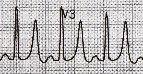
Tall T waves
T waves are tall if they are:
  • > 5mm in the limb leads and
  • > 10mm in the chest leads (the same criteria as ‘small’ QRS complexes).

Tall T waves can be associated with:
  • Hyperkalaemia (“Tall tented T waves”)
  • Hyper-acute STEMI
Peaked T
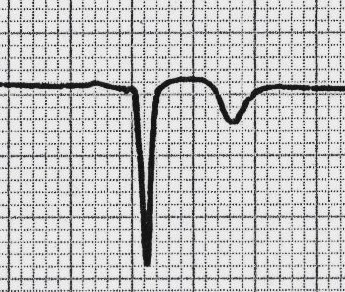
Inverted T waves
T waves are normally inverted in V1 and inversion in lead III is a normal variant.
Inverted T waves in other leads are a nonspecific sign of a wide variety of conditions:
  • Ischaemia
  • Bundle branch blocks (V4 – 6 in LBBB and V1 – V3 in RBBB)
  • PE
  • LVH (in the lateral leads)
  • HCM (widespread)
  • General illness
Around 50% of ITU admissions have some evidence of T wave inversion during their stay.
Comment on the distribution of the T wave inversion e.g. anterior / lateral / posterior leads.
You must take this ECG finding & apply it in the context of your patient.
TWI
Biphasic T waves
Biphasic T waves have two peaks and can be indicative of ischaemia and hypokalaemia.

Flattened T waves
Another non-specific sign, this may represent ischaemia or electrolyte imbalance.

U waves
Not a common finding.
The U wave is a > 0.5mm deflection after the T wave best seen in V2 or V3.
These become larger the slower the bradycardia – classically U waves are seen in various electrolyte imbalances or hypothermia, or antiarrhythmic therapy (such as digoxin, procainamide or amiodarone).
Prominent U waves in a patient with Hypokalaemia
Prominent U waves in a patient with Hypokalaemia 5
Summary
Having a system whilst working through ECGs is essential until you gain the experience required to start using pattern recognition to speed up the process.

Saturday, March 12, 2016

Gut-Microbial Role in Heart Disease!

The new study published in the journal Cell shows that TMAO (produced by gut microbes from degradation of meat, egg yolk etc) directly alters platelet function, increasing thrombosis (blood clot) potential, which could potentially be the mechanism by which TMAO increases heart attack and stroke risk.

Wednesday, February 3, 2016

For Women Thinking about Hormone Replacement:

Progesterone; YES. Progestins: NO
We start with A Fine Current Survey by a Physician which clearly restates what we have been saying now for years (OMG it's that long already 
smile emoticon
However we must first look at how Progesterone metabolites are key both to Brain and Breast and how that key role is based ultimately on the advent of new cells from progenitors every day in both tissues and how Progesterone's metabolites are key to growth of breast tissue and to neurogenesis.
The confusion between Progesterone and synthetic Progestins is extremely damaging to those patients whose physicians are unaware of the fact that Progesterone Metabolites are necessary for the proper functioning of both Brain and Breast.
These metabolites, one of them being" allopregnanolone", must be in balance for brain function and breast function to continue properly, MORE IMPORTANT, as we with the advent of researh in all areas of stem cell progress, "Function" in all tissues and organs is related to "genesis" of new cells from progenitors, whether it is neurogenesis in the brain or growth of breast tissue from progenitors.
When progestins are administered they DO NOT LEAD to the production of the same Progesterone metabolites as the "real thing". In fact, they subvert the natural and needed balance that is provided by these metabolites in both the Brain and the Breast.
Down below we simply cite what the medical advice there is (because we very much agree with it)
However, what is not stressed in the clinical advice below and that we will stress here is that progestins unlike bio-identical progesterone operate very differently in the brain as well as in the body of the woman taking them. Down further below we will appreciate that they also act very differently in the breast for the same reasons
Progesterone is essential within the brain in many ways, but one key aspect of progesterone is the crucial role its metabolites, including, in particular, allopregnanolone, are essential for the maintenance of emotional stability and avoiding anxiety based disruption with both mood and attention. Progestins do not lead to the production of these metabolites and therefore are corrosive of the mind and the spirit.
Here is an interesting review study on how vital allopregnanolone, one of the key metabolites is to determining "mood" and indeed in all Brain function
The role of Allopregnanolone in Depression and Anxiety.
http://www.ncbi.nlm.nih.gov/pubmed/24215796

The authors state:
"Neuroactive steroids such as allopregnanolone do not only act as transcriptional factors in the regulation of gene expression after intracellular back-oxidation into the 5-α pregnane steroids but may also alter neuronal excitability through interactions with specific neurotransmitter receptors.
In particular, certain 3α-reduced metabolites of progesterone such as 3α,5α-tetrahydroprogesterone (allopregnanolone) and 3α,5β-tetrahydroprogesterone (pregnanolone) are potent positive allosteric modulators of the GABA(A) receptor complex.
During the last years, the downregulation of neurosteroid biosynthesis has been intensively discussed to be a possible contributor to the development of anxiety and depressive disorder.
Reduced levels of allopregnanolone in the peripheral blood or cerebrospinal fluid were found to be associated with major depression, anxiety disorders, premenstrual dysphoric disorder, negative symptoms in schizophrenia, or impulsive aggression.
The importance of allopregnanolone for the regulation of emotion and its therapeutical use in depression and anxiety may not only involve GABAergic mechanisms, but probably also includes enhancement of neurogenesis, myelination, neuroprotection, and regulatory effects on HPA axis function.
Certain pharmacokinetic obstacles limit the therapeutic use of natural neurosteroids (low bioavailability, oxidation to the ketone). Until now synthetic neuroactive steroids could not be established in the treatment of anxiety disorders or depression. However, the translocator protein (18 kDa) (TSPO) which is important for neurosteroidogenesis has been identified as a potential novel target. TSPO ligands such as XBD 173 increase neurosteroidogenesis and have anxiolytic effects with a favorable side effect profile.
If we look further into the events with the breast we will likely find the same massively important and vastly different effects, of Progesterone versus synthetic progestins, on the genes that determine the course of progenitor cell development in the breast and surely that will also help account for the differential in breast cancer risk
What leads the Progestins to increase risk of Breast Cancer is very much related to the same disparity in yield of Progesterone metabolites that causes Progestins to cause Depression, Anxiety and Mood disorders in the Brain.
The authors state below, what we must suspect if we know how the body works. Nature does not invent a new "wheel' for every tissue and organ in the body. It just tweaks what is has to work with....just a bit.
Progesterone metabolites in breast cancer.

"In the past, the actions of the Progesterone metabolizing enzymes generally have been equated to a means of reducing the P concentration in the tissue microenvironment, and the products have been dismissed as inactive waste metabolites.
In human breast tissues and cell lines, the following P-metabolizing enzymes have been identified: 5α-reductase, 3α-hydroxysteroid oxidoreductase (3α-HSO), 3β-HSO, 20α-HSO, and 6α-hydroxylase.
Rather than providing diverse pathways for inactivating and controlling the concentration of P in breast tissue microenvironments, it is proposed that the enzymes act directly on P to produce two types of autocrines/paracrines with opposing regulatory roles in breast cancer.
In vitro studies on a number of breast cell lines indicate that 3αHP promotes normalcy by downregulating cell proliferation and detachment, whereas 5αP promotes mitogenesis and metastasis by stimulating cell proliferation and detachment. The hormones bind to novel, separate, and specific plasma membrane-based receptors and influence opposing actions on mitosis, apoptosis, and cytoskeletal and adhesion plaque molecules via cell signaling pathways.
The evidence suggests that the promotion of breast cancer is related to changes in in situ concentrations of cancer-inhibiting and -promoting P metabolites."
We can only guess at what little shop of horrors there is awaitng them in the brains and breastsof women who utilize contraceptives with progestins as their pivotal ingredient. The pharmaceutical industry surely does not want to explore that...and likely never will...and indeed will never allow it.
Because of the confusion by the Women's Health Initiative Study at the beginning of this century between Progesterone and Progestins, women continue to be afraid of progesterone, thinking it is the same as progestin. Don’t be confused here.
The physician's clinical advice with which we began this post continues:
"You can see that if most physicians and lay people believe progestins to be equivalent to progesterone, they misinterpret these studies to think progesterone is harmful and can promote breast cancer. Progesterone doesn’t do this. Progestins are not the same as progesterone.
Progesterone is a hormone the female body makes and needs for many health reasons. However, synthetic chemicals (progestins like Provera®) do not have the same benefits. It is vital that women and practicing physicians understand that bio-identical progesterone boosts wellness, and progestins are risky.
We also know that progesterone is beneficially active upon breast tissue. In fact, progesterone reduces estrogen’s stimulation of breast cancer growth. This was described by the authority in modern gynecological endocrinology, Dr. Leon Speroff, who noted, “Evidence indicates that with increasing duration of exposure, progesterone can limit breast epithelial growth as it does with endometrial epithelium
What’s more, the studies all show that while progesterone lowers breast cancer risk, progestins (synthetic forms of progesterone) do not. It is vital to know the difference between these two if you are considering progesterone hormone supplementation. The alleged “experts” from the American College of Gynecology love to denigrate “so-called” bio-identical progesterone while promoting the use of synthetic progestins.
This doesn’t account for the fact that synthetic progestins promote breast cancer and heart disease, while progesterone beneficially lowers breast cancer and heart disease risk.
The Women’s Health Initiative reported some startling information in 2002. It revealed that even though some 50 million women had been prescribed synthetic oral estrogen plus progestin therapy for better health (in the belief it would lower heart disease and breast cancer risk) since 1972, it was a mistake.
Prescribing artificial hormones was really an experiment. Nobody knew the long-term effect of these synthetic chemicals. The study reported in the Journal of the American Medical Association (JAMA) showed that Premarin® (a synthetic estrogen) and Provera® (a synthetic progestin) users had increased rates of heart disease and breast cancer, not lower rates.
Here we include a review of the horrors inflicting on peri-menopausal and post menopausal women by that Women's Heath Initative of 2002 ( and really additionally by the politically correct apologists for that bogus study who, over the past 15 years, have politcally correctly and tactfully not spoken out strongly enough on how totally incompetent were the researchers who perfomed that study and how damaging were the consequences of its misleading findings).
We put this historical essay below together for our "Gender Research for All Genders" group on Facebook, but the data and the foolishness where all based on studies of menopausal women.
https://www.academia.edu/…/Your_Heart_and_Estradiol_Beyond_…
One other important difference between progestins and bio-identical progesterone is their influence on cardiovascular health. Progestins cause potentially harmful vasoconstriction (blood vessels narrow); progesterone stimulates vascular relaxation. Also, lipid profiles (blood fats) are worsened with progestins, but improved with progesterone. There are plenty of studies to show this in the peer-reviewed scientific literature, too.
The enzymes needed to metabolize progesterone properly into either 11-deoxycorticosterone or 17-hydroxyprogesterone requires enzymes that we know the human body has. However, with progestins, we do not know if there are enzymes that will convert it to something safe or to something that is unsafe over time
The synthetic hormone problem continues and younger women still receive synthetic progestins for reasons not relating to birth control. Doctors are not acknowledging that the oral contraceptive “pill” (aka OCP) has a link to breast cancer in later years, too. Most studies of OCP and new breast cancers in women before age 40 (the worst ones) show a definite link between cancer and the use of the OCP for long durations.

Thursday, December 17, 2015

What high blood pressure (hypertension) can do


High blood pressure is a major cause of heart disease and strokes, but it also cause kidney disease, some types of dementia and eye problems.
Your brain
High blood pressure is one of the leading causes of strokes, and can lead to a form of dementia called vascular dementia:
Your heart and arteries
High blood pressure can severely damage your arteries and blood vessels. It can also cause heart disease and enlargement of the heart muscle, which can be very dangerous to your health:
Your kidneys
Your kidneys have a very important role in removing waste products from your body. High blood pressure can damage your kidneys and, in addition, damage to your kidneys can raise your blood pressure:
Your eyes and limbs
High blood pressure does not just affect your internal organs. It can also damage the blood vessels in your eyes and limbs, causing sight and mobility problems:
control high blood pressure without medication
1. Blood pressure often increases as weight increases. Being overweight also can cause disrupted breathing while you sleep (sleep apnea), which further raises your blood pressure.
2. Regular physical activity — at least 30 minutes most days of the week — can lower your blood pressure by 4 to 9 millimeters of mercury (mm Hg). It's important to be consistent because if you stop exercising, your blood pressure can rise again.
3. Eating a diet that is rich in whole grains, fruits, vegetables and low-fat dairy products and skimps on saturated fat and cholesterol can lower your blood pressure by up to 14 mm Hg. This eating plan is known as the Dietary Approaches to Stop Hypertension (DASH) diet.
4. Reduce sodium in your diet
Even a small reduction in the sodium in your diet can reduce blood pressure by 2 to 8 mm Hg.
இரத்த அழுத்தம் என்றால் என்ன
உடலில் உள்ள திசுக்களுக்குத் தேவையான உணவும் பிராண வாயும் எப்பொழுதும் கிடைக்கச் செய்யவும், அந்த திசுக்கள் உண்டாக்கும் கழிவுப் பொருட்களை வெளியேறச் செய்யவும் ஒரு அமைப்பு நம் உடலுக்குத் தேவைப்படுகிறது. இந்த அமைப்புதான் இரத்த ஓட்டம் என்பது. இந்த இரத்த ஓட்டம் ஒருவித அழுத்தத்தினால்தான் ஓடிக்கொண்டு இருக்கிறது. இதற்குத்தான் இரத்த அழுத்தம் என்று (Blood pressure) பெயர் .
உயர் இரத்த அழுத்தம் உண்டாவதற்கான காரணங்கள்
உயர் இரத்த அழுத்தத்திற்கு எந்தக் காரணமும் கண்டுபிடிக்க முடியாத நிலையில் அதற்கு முதல் நிலை உயர் இரத்த அழுத்தம் (Essential hypertension) என்று பெயர். இதில் பரம்பரை மரபு அணுகோளாறும் அடங்கும். மற்ற காரணங்களால் உண்டாகும் உயர் ரத்த அழுத்தத்திற்கு இரண்டாவது நிலை உயர் இரத்த அழுத்தம் (Secondary hypertension) என்று பெயர்.

Wednesday, November 11, 2015

Diabetes drug reduces risk of heart failure and may prevent it

In the trial, patients with type 2 diabetes and risk factors for heart disease were randomized to receive once-daily doses of either the glucose-lowering drug empagliflozin (10 mg or 25 mg doses), or a placebo. The drug or placebo was given in addition to standard care.
At the end of the trial period, investigators found that patients treated with the drug experienced reductions in blood sugar and blood pressure, as well as weight loss, compared to those on placebo.

Friday, October 30, 2015

Eating more fruits, veggies in youth linked to healthy heart decades later


Eating more fruits and vegetables as a young adult may keep your arteries free of heart disease 20 years later, according to research in the American Heart Association journal Circulation.
Researchers found that eating more fruits and vegetables as young adults was associated 26 percent lower odds of developing calcified coronary artery plaque 20 years later. Coronary artery calcium can be measured bya CT scan to detect the presence and amount of atherosclerosis, a disease that hardens arteries and underlies many types of heart disease.

Previous studies have shown a strong association between eating more fruits and vegetables and reduction in heart disease risk among middle-age adults. However, this is the first study to examine whether eating more fruits and vegetables as young adults could produce a measurable improvement in the health of their heart and blood vessels years later.
http://sciencemission.com/site/index.php…

Sunday, October 4, 2015

Captopril ( ACE Inhibitor ) ::


Captopril is an ACE inhibitor. ACE stands for Angiotensin Converting Enzyme. Captopril is used to treat high blood pressure (hypertension), congestive heart failure, kidney problems caused by diabetes, and to improve survival after a heart attack.
Indication
1. Hypertension
2. Heart Failure
3. Left Ventricular Dysfunction After Myocardial Infarction
4. Diabetic Nephropathy

Side Effects
1. Cough
2. Flushing (warmth, redness, or tingly feeling)
3. Numbness, tingling, or burning pain in your hands or feet;
4. Loss of taste sensation
5. Mild skin itching or rash.
6. Stomach pain
7. Difficulty breathing

Monday, August 24, 2015

Acute Coronary Syndrome (ACS) ::


Acute Coronary Syndrome (ACS) is a term given to various heart conditions including a Heart Attack (Myocardial Infarction) and Unstable Angina. These conditions are due to there being a reduced amount of blood flowing to a part of the heart. Various treatments are given and these usually depend on the type of ACS. Treatments help to ease the pain, improve the blood flow and to prevent any future complications.
Myocardial Infarction : 
If you have a myocardial infarction, a coronary artery or one of its smaller branches is suddenly blocked. The part of the heart muscle supplied by this artery loses its blood (and oxygen) supply. This part of the heart muscle is at risk of dying unless the blockage is quickly undone. (The word infarction means death of some tissue due to a blocked artery which stops blood from getting past.) In addition to being known as a heart attack, a myocardial infarction is sometimes called a coronary thrombosis.

Unstable angina :
Unstable angina occurs when the blood clot causes a reduced blood flow, but not a total blockage. This means that the heart muscle supplied by the affected artery does not die (infarct).
Causes
Acute coronary syndrome is most often a complication of plaque buildup in the arteries in your heart (coronary atherosclerosis) These plaques, made up of fatty deposits, cause the arteries to narrow and make it more difficult for blood to flow through them.
Eventually, this buildup means that your heart can't pump enough oxygen-rich blood to the rest of your body, causing chest pain (angina) or a heart attack. Most cases of acute coronary syndrome occur when the surface of the plaque buildup in your heart arteries ruptures and causes a blood clot to form. The combination of the plaque buildup and the blood clot dramatically limits the amount of blood flowing to your heart muscle. If the blood flow is severely limited, a heart attack will occur.
Various other uncommon conditions can also block a coronary artery. For example :
- Inflammation of the coronary arteries (rare).
- A stab wound to the heart.
- A blood clot forming elsewhere in the body (for example, in a heart chamber) and travelling to a coronary artery where it gets stuck.
- Taking cocaine, which can cause a coronary artery to go into spasm.
- Complications from heart surgery.
Sign & Symptoms
1. The most common symptom of a ACS is having severe Chest Pain. The pain often feels like a heavy pressure on your chest. The pain may also travel up into your jaw and down your left arm, or down both arms.
2. Nausea / Vomiting
3. Shortness of Breath (dyspnea)
4. Sudden, heavy sweating (diaphoresis)
5. Feel sick and feel faint
6. Abdominal Pain
7. Pain similar to heartburn
8. Clammy skin
9. Lightheadedness
10. Dizziness or fainting
See More >>>>>
http://healthmedicalinfohmi.blogspot.com/…/acute-coronary-s…

Thursday, August 20, 2015

WHAT IS HEART MURMUR

This is an unexpected sound that a doctor hears when they listen to the beating of someone’s heart through a stethoscope. They can hear the normal sounds made by the blood flowing through the heart, and of the heart valves opening and closing. If there is alteration to the smooth flow of blood through the heart this can be heard as a ‘murmur’. These can be heard when the heart relaxes – knownas a ‘diastolic’ murmur – or when it contracts, calleda ‘systolic’ murmur.

¤ CAUSES
Many murmurs are completely harmless – pregnant women often have them because of the increased amount of blood in their circulation for example – and the heart can be totally healthy and still have a murmur, such as sometimes occurs in people with anaemia. This means it is quite possible to have a heart murmur and require no treatment.
Sometimes murmurs are caused by congenital conditions such as a hole in the heart, or if there are problems with the heart valves such as prolapsed, leaking or narrowed valves.
About 30% of children will have a heart murmur at some time, but the vast majority of these are harmless (or ‘innocent’), disappearing as the child gets older or by the time they reach puberty.
Common conditions can make your heart beat faster and lead to heart murmurs. It can happen if you're pregnant or if you have:
  • Anemia
  • High blood pressure
  • Overactive thyroid
  • Fever
It could also be a problem with a heart valve. The valves close and open to let blood flow through the heart's two upper chambers (the atria) and two lower chambers (the ventricles). Valve problems include:
Mitral valve prolapse: Normally, your mitral valve closes completely when the lower left chamber of your heart contracts. It stops blood from flowing back into the upper left chamber. If part of that valve balloons out so that it doesn't close properly, you have mitral valve prolapse. This causes a clicking sound as your heart beats. It's fairly common and it's often not serious. But it can lead to the blood flowing backward through the valve, also called regurgitation.
Mitral valve or aortic stenosis: Your mitral or aortic valves are on the left side of your heart. If they narrow, which doctors call stenosis, your heart must work harder to pump blood out to the rest of your body. If untreated, it can wear out your heart and can lead to heart failure. You might be born with this condition. It can also happen as part of aging, or as a result of scarring from infections, such as rheumatic fever.
Aortic sclerosis and stenosis: One in three elderly people have a heart murmur due to the scarring, thickening, or stiffening (which doctors call sclerosis) of the aortic valve, although it hasn't narrowed. It's usually not dangerous, since the valve can work for years after the murmur starts. Aortic sclerosis is usually seen in people who have heart disease. But over time, the valve can narrow, also called stenosis. This can lead to chest pain, shortness of breath, or passing out. In some cases, the valve may need to be replaced.
Mitral or aortic regurgitation: In this case, regurgitation means the blood is going the wrong way through the mitral valve or the aortic valve. To counteract this backflow, the heart must work harder to force blood through the damaged valve. Over time, this can weaken or enlarge the heart and can lead to heart failure.
Congenital heart defects: About 25,000 babies are born each year with heart defects, such as holes in heart walls or abnormal heart valves. Surgery can correct many of these problems.

Treatment

Many children and adults have harmless heart murmurs, which don't need treatment.
If another condition, such as high blood pressure, is causing your heart murmurs, your doctor will treat the underlying cause.
Some types of heart valve disease may require medication or surgery:
  • Medicines to prevent blood clots, control irregular heartbeat or palpitations, and lower blood pressure
  • Diuretics to remove excess salt and water from the body, making it easier for your heart to pump
  • Surgery to correct heart defects someone is born with
  • Surgery to correct certain types of heart valve disease
  • It's not common, but doctors sometimes ask people to takeantibiotics to help prevent heart infection before dental work or some kinds of surgery.

Diagnosis

Usually, doctors find heart murmurs during a physical exam. Your doctor will be able to hear your heart murmur when listening to your heart with a stethoscope.
Your doctor may order one or more of the following tests to see whether your heart murmur is innocent or whether it is caused by acquired valve disease or a defect you were born with:
  • Electrocardiogram (ECG), which measures the electrical activity of the heart
  • Chest X-rays to see if the heart is enlarged due to heart or valve disease
  • Echocardiography, which uses sound waves to map the heart's structure

What About Prevention?

In most cases, you can't prevent heart murmurs. The exception: Treating an underlying condition, such as high blood pressure, or avoiding heart valve infection, can stop heart murmurs before they start.

When to Call Your Doctor

Call your doctor about a heart murmur if you feel:
  • Chest pain
  • Breathlessness, fatigue, or fainting for no obvious reason
  • Heart palpitations

☞ Tejin Rana.