Search This Blog

Showing posts with label Anatomy and Physiology. Show all posts

Monday, June 20, 2016

Inside the cell

This is an incredible tripthat we can only go through an animation that I find extraordinary. The first time I saw this video, I was stunned by how they could tell a cell so just in its complexity. I think that will appeal to each of you because it shows different mechanisms that, even as you read these lines, are happening constantly.
I believe that the graphic representation of the ribosome and how they work is fantastic, not least of all the rest.
"In a cell, the starting point of a more complex realization that call an organism."

Friday, October 30, 2015

SCAPULAR BLADE FRACTURE

The shoulder blade (scapula) is a triangular-shaped bone that is protected by a complex system of surrounding muscles. Scapula fractures represent less than 1% of all broken bones and many of them can be treated without surgery.
High-energy, blunt trauma injuries, such as those experienced in a motorcycle or motor vehicle collision or falling from a significant height, can cause a scapula fracture. Other major injuries often accompany scapular fractures, such as fractures in the shoulder, collarbone and ribs, or damage to the head, lungs, or spinal cord.
One or more parts of the scapula may be fractured.
  • Scapular body (50% to 60% of patients)
  • Scapular neck (25% of patients)
  • Glenoid
  • Acromion
  • Coracoid
Symptoms
The most common symptoms of a scapula fracture include:
  • Extreme pain when you move the arm
  • Swelling around the back of the shoulder
  • Scrapes around the affected area.

Monday, June 8, 2015

Menstrual Cycle ::


The menstrual cycle is the cycle of natural changes that occurs in the uterus and ovary as an essential part of making sexual reproduction possible. It is also known as Menstruation, Menses, Period. In humans, the length of a menstrual cycle varies greatly among women (ranging from 21 to 35 days), with 28 days designated as the average length. Each cycle can be divided into three phases based on events in the ovary (ovarian cycle) or in the uterus (uterine cycle).
The ovarian cycle consists of the follicular phase, ovulation, and luteal phase whereas the uterine cycle is divided into menstruation, proliferative phase, and secretory phase. Both cycles are controlled by the endocrine system and the normal hormonal changes that occur can be interfered with using hormonal contraception to prevent reproduction.
The menstrual cycle can be described by the ovarian or uterine cycle. The ovarian cycle describes changes that occur in the follicles of the ovary whereas the uterine cycle describes changes in the endometrial lining of the uterus. Both cycles can be divided into three phases. The ovarian cycle consists of the follicular phase, ovulation, and the luteal phase whereas the uterine cycle consists of menstruation, proliferative phase, and secretory phase.
The three phases of Menstrual cycle are :
1. Menstrual Period
✔ On Day 1 of your cycle, the thickened lining (endometrium) of the uterus begins to shed. You know this as menstrual bleeding from the vagina. A normal menstrual period can last 4 to 6 days.
✔ Most of your menstrual blood loss happens during the first 3 days.
✔ This is also when you might have cramping pain in your pelvis, legs, and back. Cramps can range from mild to severe. The cramping is your uterus contracting, helping the endometrium shed.
✔ In general, any premenstrual symptoms that you've felt before your period will go away during these first days of your cycle.
See More [ 2. Follicular Phase, 3. Luteal (premenstrual) Phase ]
Premenstrual Syndrome (PMS) ::
Premenstrual Syndrome (PMS) has a wide variety of symptoms, including mood swings, tender breasts, food cravings, crying, oversensitivity, fatigue, irritability and depression. It is also known as PMS or Premenstrual Tension (PMT) or Premenstrual Dysphoric Disorder (PMDD). PMS symptoms occur 1 to 2 weeks before menstrual period (menstruation) starts. PMS can affect menstruating women of any age and the effect is different for each woman.
Causes
✿ Cyclic changes in hormones.
✿ Chemical changes in the brain.
✿ Depression (Feeling of sadness for periods of at least 2 weeks).
Sign & Symptoms
Symptoms vary from woman to woman. PMS often includes both physical and emotional symptoms, such as :
1. Acne
2. Swollen or tender breasts
3. Feeling tired
4. Trouble sleeping
5. Upset stomach, bloating, constipation, or diarrhea
6. Headache or backache
7. Appetite changes or food cravings
8. Joint or muscle pain
9. Trouble with concentration or memory
10. Tension, irritability, mood swings, or crying spells
11. Anxiety or depression
top 11 tips to maintain this prolem or hygiene during your periods
1. Choose your method of sanitation :
There are a number of ways including the use of sanitary napkins, tampons and menstrual cups to stay clean. So, try to use best, comfortable sanitary napkins, pads.

2. Change regularly :
If sanitary napkin is unchange then more chance to Urinary Tract Infection (UTI), vaginal infections and skin rashes. The standard time to change a sanitary pad is once every six hours, while for a tampon is once every two hours.

3. Wash yourself regularly :
When you menstruate, the blood tends to enter tiny spaces like the skin between your labia or crust around the opening of the vagina and you should always wash this excess blood away. This practice also tends to beat bad odour from the vaginal region.

4. Right washing technique :
Always wash or clean the area in a motion that is from the vagina to the anus. Never wash in the opposite direction. Washing in the opposite direction can cause Bacteria from the anus to lodge in the vagina and urethral opening, leading to infections.
5. Have a bath regularly :
Bathing not only cleanses your body but also gives you a chance to clean your private parts well. It also helps relieve menstrual cramps, backaches, helps improve your mood and makes you feel less bloated. To get some relief from backaches and menstrual cramps, just stand under a shower of warm water that is targeted towards your back or abdomen.

6. Discard your used sanitary product properly :
It is essential to discard your used napkins or tampons properly because they are capable of spreading infections, will smell very foul. Wrapping it well before discarding it ensures that the smell and infection is contained.

7. Reduce salt and caffeine :
Avoid processed and fast foods, with their high-salt composition, and you'll help hold water retention at bay and minimize bloating. Caffeine -- found in your daily ration of coffee, tea or soda -- can irritate the stomach by increasing gastrointestinal acidity, making your cramps worse and adding to the discomfort.

8. Get enough calcium :
Foods rich in calcium -- broccoli, yogurt, cabbage and milk -- will naturally fight muscle spasms and help you avoid cramps. Women need at least 1,200 mg of calcium daily, and choosing to get yours though leafy green veggies like kale and spinach has the added advantage of an extra dose of vitamins that can help lighten your flow.

9. Exercise :
Stretch those abdominal muscles with a gentle yoga workout. While you may not feel energetic enough for your normal fitness routine, increasing the blood flow to your pelvic region will give quick relief from cramps.

10. Use of Medicine :
Menstrual pain and cramps can be reduced by drug treatment with Ibuprofen, Aspirin or Naproxen. If you have a regular menstrual cycle, taking one of these pills one day before your period starts can often get rid of the normal discomfort.

11. Apply some Heat :
Stretch out and relax for a few minutes with a heating pad set on low resting on your abdomen. The heat will loosen tight muscles and ease cramping, while the quiet time lowers anxiety and allows you to recharge and feel less irritated and moody.

References
1. http://www.sheknows.com/health-and-wellness
2. http://www.thehealthsite.com/

Friday, January 30, 2015

IMPORTANT LANDMARKS IN FETAL DEVELOPMENT ::


1. Fetus starts swallowing by 10-12 weeks.
2. Fetal breathing movts being by 11 weeks.
3. Fetus starts producing urine by 12 weeks.
4. Ovaries and testis are 1st recognised by 8 weeks.
5. External genitalia are first recognised by 12 weeks.
6. On USG sex be determined by 14 weeks.
7. Placental circulation is established by 17-21 days after fertilisation.
8. Insulin secretion in fetus begins by 12 weeks.

Tuesday, November 18, 2014

Anatomy of Human Brain Surface

The brain is one of the most complex and magnificent organs in the human body. Our brain gives us awareness of ourselves and of our environment, processing a constant stream of sensory data. It controls our muscle movements, the secretions of our glands, and even our breathing and internal temperature. Every creative thought, feeling, and plan is developed by our brain. The brain’s neurons record the memory of every event in our lives.
In fact, the human brain is so complicated that it remains an exciting frontier in the study of the body; doctors, psychologists, and scientists are continually endeavoring to learn exactly how the many structures of the brain work together intricately to create our powerful human mind.

Anatomy of the Brain

There are different ways of dividing the brain anatomically into regions. Let’s use a common method and divide the brain into three main regions based on embryonic development: the forebrain, midbrain and hindbrain. Under these divisions:
  • The forebrain (or prosencephalon) is made up of our incredible cerebrum, thalamus, hypothalamus and pineal gland among other features. Neuroanatomists call the cerebral area the telencephalon and use the term diencephalon (or interbrain) to refer to the area where our thalamus, hypothalamus and pineal gland reside.
  • The midbrain (or mesencephalon), located near the very center of the brain between the interbrain and the hindbrain, is composed of a portion of the brainstem.
  • The hindbrain (or rhombencephalon) consists of the remaining brainstem as well as our cerebellum and pons. Neuroanatomists have a word to describe the brainstem sub-region of our hindbrain, calling it the myelencephalon, while they use the wordmetencephalon in reference to our cerebellum and pons collectively.
Before exploring these different regions of the brain, first let’s define the important types of cells and tissues that are the building blocks of them all.
Histology
Brain cells can be broken into two groups: neurons and neuroglia.
Neurons, or nerve cells, are the cells that perform all of the communication and processing within the brain. Sensory neurons entering the brain from the peripheral nervous system deliver information about the condition of the body and its surroundings. Most of the neurons in the brain’s gray matter are interneurons, which are responsible for integrating and processing information delivered to the brain by sensory neurons. Interneurons send signals to motor neurons, which carry signals to muscles and glands.
Neuroglia, or glial cells, act as the helper cells of the brain; they support and protect the neurons. In the brain there are four types of glial cells: astrocytes, oligodendrocytes, microglia, and ependymal cells.
  • Astrocytes protect neurons by filtering nutrients out of the blood and preventing chemicals and pathogens from leaving the capillaries of the brain.
  • Oligodendrocytes wrap the axons of neurons in the brain to produce the insulation known as myelin. Myelinated axons transmit nerve signals much faster than unmyelinated axons, so oligodendrocytes accelerate the communication speed of the brain.
  • Microglia act much like white blood cells by attacking and destroying pathogens that invade the brain.
  • Ependymal cells line the capillaries of the choroid plexuses and filter blood plasma to produce cerebrospinal fluid.
The tissue of the brain can be broken down into two major classes: gray matter and white matter.
  • Gray matter is made of mostly unmyelinated neurons, most of which are interneurons. The gray matter regions are the areas of nerve connections and processing.
  • White matter is made of mostly myelinated neurons that connect the regions of gray matter to each other and to the rest of the body. Myelinated neurons transmit nerve signals much faster than unmyelinated axons do. The white matter acts as the information highway of the brain to speed the connections between distant parts of the brain and body.
Now let’s begin exploring the main structures of our awesome human brain.
HINDBRAIN (RHOMBENCEPHALON)

Brainstem
Connecting the brain to the spinal cord, the brainstem is the most inferior portion of our brain. Many of the most basic survival functions of the brain are controlled by the brainstem.
The brainstem is made of three regions: the medulla oblongata, the pons, and the midbrain. A net-like structure of mixed gray and white matter known as the reticular formation is found in all three regions of the brainstem. The reticular formation controls muscle tone in the body and acts as the switch between consciousness and sleep in the brain.
The medulla oblongata is a roughly cylindrical mass of nervous tissue that connects to the spinal cord on its inferior border and to the pons on its superior border. The medulla contains mostly white matter that carries nerve signals ascending into the brain and descending into the spinal cord. Within the medulla are several regions of gray matter that process involuntary body functions related to homeostasis. The cardiovascular center of the medulla monitors blood pressure and oxygen levels and regulates heart rate to provide sufficient oxygen supplies to the body’s tissues. The medullary rhythmicity center controls the rate of breathing to provide oxygen to the body. Vomiting, sneezing, coughing, and swallowing reflexes are coordinated in this region of the brain as well.
The pons is the region of the brainstem found superior to the medulla oblongata, inferior to the midbrain, and anterior to the cerebellum. Together with the cerebellum, it forms what is called the metencephalon. About an inch long and somewhat larger and wider than the medulla, the pons acts as the bridge for nerve signals traveling to and from the cerebellum and carries signals between the superior regions of the brain and the medulla and spinal cord.
Cerebellum
The cerebellum is a wrinkled, hemispherical region of the brain located posterior to the brainstem and inferior to the cerebrum. The outer layer of the cerebellum, known as the cerebellar cortex, is made of tightly folded gray matter that provides the processing power of the cerebellum. Deep to the cerebellar cortex is a tree-shaped layer of white matter called the arbor vitae, which means ‘tree of life’. The arbor vitae connects the processing regions of cerebellar cortex to the rest of the brain and body.
The cerebellum helps to control motor functions such as balance, posture, and coordination of complex muscle activities. The cerebellum receives sensory inputs from the muscles and joints of the body and uses this information to keep the body balanced and to maintain posture. The cerebellum also controls the timing and finesse of complex motor actions such as walking, writing, and speech.
MIDBRAIN (MESENCEPHALON)

The midbrain, also known as the mesencephalon, is the most superior region of the brainstem. Found between the pons and the diencephalon, the midbrain can be further subdivided into 2 main regions: the tectum and the cerebral peduncles.
  • The tectum is the posterior region of the midbrain, containing relays for reflexes that involve auditory and visual information. The pupillary reflex (adjustment for light intensity), accommodation reflex (focus on near or far away objects), and startle reflexes are among the many reflexes relayed through this region.
  • Forming the anterior region of the midbrain, the cerebral peduncles contain many nerve tracts and the substantia nigra. Nerve tracts passing through the cerebral peduncles connect regions of the cerebrum and thalamus to the spinal cord and lower regions of the brainstem. The substantia nigra is a region of dark melanin-containing neurons that is involved in the inhibition of movement. Degeneration of the substantia nigra leads to a loss of motor control known as Parkinson’s disease.
FOREBRAIN (PROSENCEPHALON)

Diencephalon
Superior and anterior to the midbrain is the region known as the interbrain, or diencephalon. The thalamus, hypothalamus, and pineal glands make up the major regions of the diencephalon.
  • The thalamus consists of a pair of oval masses of gray matter inferior to the lateral ventricles and surrounding the third ventricle. Sensory neurons entering the brain from the peripheral nervous system form relays with neurons in the thalamus that continue on to the cerebral cortex. In this way the thalamus acts like the switchboard operator of the brain by routing sensory inputs to the correct regions of the cerebral cortex. The thalamus has an important role in learning by routing sensory information into processing and memory centers of the cerebrum.
  • The hypothalamus is a region of the brain located inferior to the thalamus and superior to the pituitary gland. The hypothalamus acts as the brain’s control center for body temperature, hunger, thirst, blood pressure, heart rate, and the production of hormones. In response to changes in the condition of the body detected by sensory receptors, the hypothalamus sends signals to glands, smooth muscles, and the heart to counteract these changes. For example, in response to increases in body temperature, the hypothalamus stimulates the secretion of sweat by sweat glands in the skin. The hypothalamus also sends signals to the cerebral cortex to produce the feelings of hunger and thirst when the body is lacking food or water. These signals stimulate the conscious mind to seek out food or water to correct this situation. The hypothalamus also directly controls the pituitary gland by producing hormones. Some of these hormones, such as oxytocin and antidiuretic hormone, are produced in the hypothalamus and stored in the posterior pituitary gland. Other hormones, such as releasing and inhibiting hormones, are secreted into the blood to stimulate or inhibit hormone production in the anterior pituitary gland.
  • The pineal gland is a small gland located posterior to the thalamus in a sub-region called the epithalamus. The pineal gland produces the hormone melatonin. Light striking the retina of the eyes sends signals to inhibit the function of the pineal gland. In the dark, the pineal gland secretes melatonin, which has a sedative effect on the brain and helps to induce sleep. This function of the pineal gland helps to explain why darkness is sleep-inducing and light tends to disturb sleep. Babies produce large amounts of melatonin, allowing them to sleep as long as 16 hours per day. The pineal gland produces less melatonin as people age, resulting in difficulty sleeping during adulthood.
Cerebrum
The largest region of the human brain, our cerebrum controls higher brain functions such as language, logic, reasoning, and creativity. The cerebrum surrounds the diencephalon and is located superior to the cerebellum and brainstem. A deep furrow known as thelongitudinal fissure runs midsagittally down the center of the cerebrum, dividing the cerebrum into the left and right hemispheres. Each hemisphere can be further divided into 4 lobes: frontalparietaltemporal, and occipital. The lobes are named for the skull bones that cover them.
The surface of the cerebrum is a convoluted layer of gray matter known as the cerebral cortex. Most of the processing of the cerebrum takes place within the cerebral cortex. The bulges of cortex are called gyri (singular: gyrus) while the indentations are calledsulci (singular: sulcus).
Deep to the cerebral cortex is a layer of cerebral white matter. White matter contains the connections between the regions of the cerebrum as well as between the cerebrum and the rest of the body. A band of white matter called the corpus callosum connects the left and right hemispheres of the cerebrum and allows the hemispheres to communicate with each other.
Deep within the cerebral white matter are several regions of gray matter that make up the basal nuclei and the limbic system. The basal nuclei, including the globus pallidus, striatum, and subthalamic nucleus, work together with the substantia nigra of the midbrain to regulate and control muscle movements. Specifically, these regions help to control muscle tone, posture, and subconscious skeletal muscle. The limbic system is another group of deep gray matter regions, including the hippocampus and amygdala, which are involved in memory, survival, and emotions. The limbic system helps the body to react to emergency and highly emotional situations with fast, almost involuntary actions.
With so many vital functions under the control of a single incredible organ - and so many important functions carried out in its outer layers - how does our body protect the brain from damage? Our skull clearly offers quite a bit of protection, but what protects the brain from the skull itself? Read on!
Meninges
Three layers of tissue, collectively known as the meninges, surround and protect the brain and spinal cord.
  • The dura mater forms the leathery, outermost layer of the meninges. Dense irregular connective tissue made of tough collagen fibers gives the dura mater its strength. The dura mater forms a pocket around the brain and spinal cord to hold the cerebrospinal fluid and prevent mechanical damage to the soft nervous tissue. The name dura mater comes from the Latin for “tough mother,” due to its protective nature.
  • The arachnoid mater is found lining the inside of the dura mater. Much thinner and more delicate than the dura mater, it contains many thin fibers that connect the dura mater and pia mater. The name arachnoid mater comes from the Latin for “spider-like mother”, as its fibers resemble a spider web. Beneath the arachnoid mater is a fluid-filled region known as the subarachnoid space.
  • As the innermost of the meningeal layers, the pia mater rests directly on the surface of the brain and spinal cord. The pia mater’s many blood vessels provide nutrients and oxygen to the nervous tissue of the brain. The pia mater also helps to regulate the flow of materials from the bloodstream and cerebrospinal fluid into nervous tissue.
Cerebrospinal Fluid
Cerebrospinal fluid (CSF) – a clear fluid that surrounds the brain and spinal cord – provides many important functions to the central nervous system. Rather than being firmly anchored to their surrounding bones, the brain and spinal cord float within the CSF. CSF fills the subarachnoid space and exerts pressure on the outside of the brain and spinal cord. The pressure of the CSF acts as a stabilizer and shock absorber for the brain and spinal cord as they float within the hollow spaces of the skull and vertebrae. Inside of the brain, small CSF-filled cavities called ventricles expand under the pressure of CSF to lift and inflate the soft brain tissue.
Cerebrospinal fluid is produced in the brain by capillaries lined with ependymal cells known as choroid plexuses. Blood plasma passing through the capillaries is filtered by the ependymal cells and released into the subarachnoid space as CSF. The CSF contains glucose, oxygen, and ions, which it helps to distribute throughout the nervous tissue. CSF also transports waste products away from nervous tissues.
After circulating around the brain and spinal cord, CSF enters small structures known as arachnoid villi where it is reabsorbed into the bloodstream. Arachnoid villi are finger-like extensions of the arachnoid mater that pass through the dura mater and into the superior sagittal sinus. The superior sagittal sinus is a vein that runs through the longitudinal fissure of the brain and carries blood and cerebrospinal fluid from the brain back to the heart.

Physiology of the Brain

Metabolism
Despite weighing only about 3 pounds, the brain consumes as much as 20% of the oxygen and glucose taken in by the body. Nervous tissue in the brain has a very high metabolic rate due to the sheer number of decisions and processes taking place within the brain at any given time. Large volumes of blood must be constantly delivered to the brain in order to maintain proper brain function. Any interruption in the delivery of blood to the brain leads very quickly to dizziness, disorientation, and eventually unconsciousness.
Sensory
The brain receives information about the body’s condition and surroundings from all of the sensory receptors in the body. All of this information is fed into sensory areas of the brain, which put this information together to create a perception of the body’s internal and external conditions. Some of this sensory information is autonomic sensory information that tells the brain subconsciously about the condition of the body. Body temperature, heart rate, and blood pressure are all autonomic senses that the body receives. Other information is somatic sensory information that the brain is consciously aware of. Touch, sight, sound, and hearing are all examples of somatic senses.
Motor Control
Our brain directly controls almost all movement in the body. A region of the cerebral cortex known as the motor area sends signals to the skeletal muscles to produce all voluntary movements. The basal nuclei of the cerebrum and gray matter in the brainstem help to control these movements subconsciously and prevent extraneous motions that are undesired. The cerebellum helps with the timing and coordination of these movements during complex motions. Finally, smooth muscle tissue, cardiac muscle tissue, and glands are stimulated by motor outputs of the autonomic regions of the brain.
Processing
Once sensory information has entered the brain, the association areas of the brain go to work processing and analyzing this information. Sensory information is combined, evaluated, and compared to prior experiences, providing the brain with an accurate picture of its conditions. The association areas also work to develop plans of action that are sent to the brain’s motor regions in order to produce a change in the body through muscles or glands. Association areas also work to create our thoughts, plans, and personality.
Learning and Memory
The brain needs to store many different types of information that it receives from the senses and that it develops through thinking in the association areas. Information in the brain is stored in a few different ways depending on its source and how long it is needed. Our brain maintains short-term memory to keep track of the tasks in which the brain is currently engaged. Short-term memory is believed to consist of a group of neurons that stimulate each other in a loop to keep data in the brain’s memory. New information replaces the old information in short-term memory within a few seconds or minutes, unless the information gets moved to long-term memory.
Long-term memory is stored in the brain by the hippocampus. The hippocampus transfers information from short-term memory to memory-storage regions of the brain, particularly in the cerebral cortex of the temporal lobes. Memory related to motor skills (known as procedural memory) is stored by the cerebellum and basal nuclei.
Homeostasis
The brain acts as the body’s control center by maintaining the homeostasis of many diverse functions such as breathing, heart rate, body temperature, and hunger. The brainstem and the hypothalamus are the brain structures most concerned with homeostasis.
In the brainstem, the medulla oblongata contains the cardiovascular center that monitors the levels of dissolved carbon dioxide and oxygen in the blood, along with blood pressure. The cardiovascular center adjusts the heart rate and blood vessel dilation to maintain healthy levels of dissolved gases in the blood and to maintain a healthy blood pressure. The medullary rhythmicity center of the medulla monitors oxygen and carbon dioxide levels in the blood and adjusts the rate of breathing to keep these levels in balance.
The hypothalamus controls the homeostasis of body temperature, blood pressure, sleep, thirst, and hunger. Many autonomic sensory receptors for temperature, pressure, and chemicals feed into the hypothalamus. The hypothalamus processes the sensory information that it receives and sends the output to autonomic effectors in the body such as sweat glands, the heart, and the kidneys.
Sleep
While sleep may seem to be a time of rest for the brain, this organ is actually extremely active during sleep. The hypothalamus maintains the body’s 24 hour biological clock, known as the circadian clock. When the circadian clock indicates that the time for sleep has arrived, it sends signals to the reticular activating system of the brainstem to reduce its stimulation of the cerebral cortex. Reduction in the stimulation of the cerebral cortex leads to a sense of sleepiness and eventually leads to sleep.
In a state of sleep, the brain stops maintaining consciousness, reduces some of its sensitivity to sensory input, relaxes skeletal muscles, and completes many administrative functions. These administrative functions include the consolidation and storage of memory, dreaming, and development of nervous tissue.
There are two main stages of sleep: rapid eye movement (REM) and non-rapid eye movement (NREM). During REM sleep, the body becomes paralyzed while the eyes move back and forth quickly. Dreaming is common during REM sleep and it is believed that some memories are stored during this phase. NREM sleep is a period of slow eye movement or no eye movement, culminating in a deep sleep of low brain electrical activity. Dreaming during NREM sleep is rare, but memories are still processed and stored during this time.
Reflexes
A reflex is a fast, involuntary reaction to a form of internal or external stimulus. Many reflexes in the body are integrated in the brain, including the pupillary light reflex, coughing, and sneezing. Many reflexes protect the body from harm. For instance, coughing and sneezing clear the airways of the lungs. Other reflexes help the body respond to stimuli, such as adjusting the pupils to bright or dim light. All reflexes happen quickly by bypassing the control centers of the cerebral cortex and integrating in the lower regions of the brain such as the midbrain or limbic system.
Prepared by Tim Taylor, Anatomy and Physiology Instructor

Thursday, May 22, 2014

Students design artificial kidney with 3-D printing

Detail —Three-dimensional printing has garnered coverage in the popular press for its application in the custom manufacturing of tools and mechanical parts. But six School of Engineering seniors have recently taken the application of the technology into the medical field, using 3-D printing to create body parts.

Under the direction of Anson Ma, assistant professor in the Department of Chemical and Biomolecular Engineering and the Institute of Materials Science, two three-person teams of chemical engineering students were tasked with creating an artificial kidney for their Senior Design Project using 3-D printing technology. 3-D printing is an additive manufacturing method capable of creating complex parts that are otherwise impossible or extremely difficult to produce.

The students participating were: Derek Chhiv, Meaghan Sullivan, Danny Ung, Benjamin Coscia, Guleid Awale, and Ali Rogers. They are one of the first classes of students to partner with a commercial 3-D printing company, ACT Group, to create a prototype.

The challenge the teams set out to tackle is rooted in a very real problem.

The United States Renal Data System reports that, as recently as 2009, End-Stage Renal Disease (ESRD) resulted in over 90,000 deaths. Options for treatment of renal disease are essentially limited to either an organ transplant or dialysis. However, there is a limited supply of transplantable kidneys, with demand far outstripping the supply; and dialysis is expensive and is only a temporary solution.

Source: Phys Org

Monday, December 30, 2013

Anatomy of a cell ( உடல் செல்களின் உள்ளே ஒரு பயணம்)


Cells are "building blocks" of life: all living things, whether plants, animals, people, or tiny microscopic organisms, are made up of cells. Although a cell is only about 10 micrometres across (one micrometer=one millionth of a meter!), there is still amazing complexity within it.
The plasma membrane around the cell is semi-permeable, meaning that some substances are able to enter the cell through it and some are not. Plant cells and some bacteria and algae cells have a protective cell wall in addition. Although animal cells don't have a cell wall, they are protected by other cells, such as white blood cells that fight disease.
Inside the cell is a jelly-like fluid called cytoplasm that holds a cell's organelles, special structures that perform specific cell functions. Some of the main organelles within the cell are the vacuoles, mitochondria, lysosomes, ribosomes, endoplasmic reticulum, Golgi apparatus, and the cell nucleus. Think of organelles as being similar to the organs in your body: your heart, liver, and brain are all organs, performing specific functions to make your bodywork.


Most of these organelles are present in both animal and plant cells. The endoplasmic reticulum (ER) is important in the production or synthesis of cell components. Smooth ER produces lipids and membrane proteins, while rough ER (so-called because it contains protein-producing ribosomes) produces all other proteins needed by the cell. These proteins are modified by the Golgi apparatus, which also stores and packages them for exportation from the cell. (You can think of the Golgi apparatus as a kind of shipping department in the cell.)
Vacuoles are the cell's main storage units, holding food, water, or waste until it can be used or disposed of. Mitochondria are the "powerhouses" of the cell, converting nutrients to energy. Animal cells contain lysosomes that are responsible for the reactions that break down proteins, poly- and disaccharides, and some lipids. Your white blood cells use lysosomes to "eat" disease with digestive enzymes.
diagram of animal cell
The nucleus provides the "brains" for this operation--the cell would be unable to do anything without it. The nucleus contains deoxyribonucleic acid, or DNA,  which is the genetic material of life. Messenger ribonucleic acid, or RNA, is also important, as is makes a "negative" copy (like the negative of a photograph) of the DNA and takes this information outside the nucleus to the ribosomes. At the ribosomes, transfer RNA "translates" the code from the messenger RNA, allowing the ribosomes to form a protein.

Eukaryotic cells, which include animal and plant cells, have a nucleus enclosed in membrane. Prokaryotic cells such as bacteria do not have a nuclear membrane; the genetic material is just clumped in the centre of the cell instead.
Mitosis is asexual reproduction (without the union of male and female gametes) that takes place in cells. There are four stages to this process. In very simplified terms, the cell's replicated DNA separates into two sets of identical chromosomes during prophase; the chromosomes are aligned down the centre of the cell during metaphase; the duplicate chromosomes separate during anaphase; and in telophase, two identical copies--or clones--are formed out of what was one "mother" cell, each with an identical set of chromosomes.
Sexual reproduction in cells, or meiosis, involves more stages and is much more complex, resulting in a new,  the unique combination of genetic material rather than making an identical copy.


ஆச்சர்யத்தை அள்ளி தரும் உலகின் பல விஷயங்களில் இந்த உடலும் ஒன்று. உற்று பார்த்தால் உடலில் இருக்கும் பல கட்டமைப்புகள் பிரமிப்பை உண்டு பண்ணும்.
உதாரணமாக ஒரு மனிதனின் உடலில் உள்ள மொத்த DNA வை எடுத்து அந்த நூலை நேராக நீட்டி வரிசையாக வைத்தால் அது பூமியில் இருந்து சூரியன் சென்று மீண்டும் பூமி வந்தால் எவ்வளவு தூரம் இருக்குமோ..அந்..த ... தூரம்... (இருங்க அவசர படாதீங்க.. )அந்த தூரம்.. அதை விட கிட்ட தட்ட ஒரு 300 மடங்கு அதிகம் தூரம் இருக்குமாம்...( அல்லது கிட்ட தட்ட மொத்த சூரிய குடும்ப தொலைவை போல இரு மடங்கு என்று சொல்லலாம்..'எம்மாடி..' )
DNA வில் நாம் மெமரியை பதிய முடியும் எனில் ஒரே ஒரு கிராம் DNA வில் நாம் உலகின் மொத்த டேட்டா வை சேகரித்து வைக்க முடியும். இப்படி நிறைய ஆச்சர்யங்களை சொல்லி கொண்டே போகலாம்.

"செல் " படம் வரைந்து பாகம் குறித்த பள்ளி நினைவு நம் அனைவருக்கும் இருக்கும். இன்று மீண்டும் ஒரு முறை செல் என்பதை பற்றி அதில் உள்ள உறுப்புகள் பற்றி...அது செயல் படும் விதம் பற்றி கொஞ்சம் விளக்கமாக பார்க்கலாம் வாருங்கள்..

முதலில் செல் என்பது என்ன ? ஒரு கட்டிடம் செங்களால் ஆனது என்பதை போல ஒரு உயிருள்ள உடல் எனும் கட்டுமானம் செல் எனும் செங்கல் அடுக்குகளால் ஆனது.
உலகத்தில் உள்ள தாவரம் விலங்கு மனிதன் பூச்சி எல்லாமே இப்படி தான். ஆனால் எல்லா செல்களும் ஒன்றை போலவே இருக்குமா என்றால் இல்லை. தாவர செல்கள் கொஞ்சம் மாறு பட்டவை. உதாரணமாக தாவர செல்களில் cell wall என்று ஒரு மேலடுக்கு பாதுகாப்பு இருக்கும். (தாவர உடல் கடினமாக இருக்க காரணம் இது தான் ) அது எந்த விலங்கு செல்களிலும் இல்லை. சூரிய ஆற்றலை நேராக இழுத்து சக்தியாக மாற்றும் சிறப்பு அமைப்பு தாவர செல்லில் உண்டு அதுவும் விலங்கு செல்லில் இல்லை.

இப்படி வேற்றுமை இருக்கும் அதே நேரத்தில் உலகில் உள்ள அனைத்து செல்களுக்கும் பொருந்தும் படி சில ஒற்றுமைகள் இருக்கின்றன. உதாரணமாக அனைத்து செல்லிலும் cell membrane இருக்கும் (செல்களை தன்னை சுற்றி உள்ள சுற்றுசூழலில் இருந்து பிரித்து வைக்கும் கவசம் ) அடுத்ததாக sytoplasm என்ற ஒரு ஜெல் அமைப்பு இருக்கும் (செல் பூரா நிரம்பி இருப்பது இது தான். செல்களின் பாகங்கள் இதில் தான் மிதந்து கொண்டு இருக்கும் )
அப்புறம் ஜெனடிக் மெடிரியல் இருக்கும் (உதாரணம் DNA இவைகள் தான் அந்த சம்பந்த பட்ட உயிரியின் மரபு பற்றிய தகவல்களை தாங்கி இருக்கும் )


சரி இப்போதைக்கு விலங்கு செல்கள் பற்றி மட்டும் கொஞ்சம் விளக்கமாக பார்க்கலாம்.
🧬 🧬 விலங்கு செல்லில் இரண்டு வகைகள் உள்ளது. ஒன்று சிக்கலான அமைப்பு நிறைந்த மேம்பட்ட செல்கள். அதாவது நாய் பூனை எலி பூச்சி யானை மனிதன் என்று அனைத்திலும் இருக்கும் செல்கள். (தாவர செல்கள் கூட இந்த வகையில் தான் சேரும் ) இவற்றிற்கு பெயர்" Eukaryotic cell " .
இந்த வகை செல்கள் பல உள் உறுப்புகளை... செல் உறுப்புகளை கொண்டு இருக்கும் இந்த உறுப்புகளை "organells " என்று அழைக்கிறோம்.மனித உடலில் எப்படி organs இருக்கிறதோ அப்படி செல்லின் உடல் பாகங்கள் தான் ' organells '. இதை தவிர இந்த வகை செல்கள் ஒரு உட்கருவை கொண்டு இருக்கும் (nucleus ). மற்றும் மேலும் சில சிறப்பு பாகங்களை கொண்டு இருக்கும்.

இராண்டாவது வகை செல்கள் prokaryotic cells என்று அழைக்க படுகின்றன. (இரண்டு வகை செல்களில் மூத்தது இது தான்.. இதில் இருந்து தான் யூகிரியாடிக் செல்கள் பிற்காலத்தில் பரிணாமம் அடைந்தது.. )
இவைகள் முந்திய வகை அளவு சிக்கல் இல்லாதவை. நியூக்ளியஸ் எனும் உட்கருவோ அல்லது membran போர்வையால் போர்த்த பட்ட உள்ளுறுப்புகளோ இல்லாதவை . (ஆனால் இவற்றிலும் கூட மரபணு சங்கதிகள் மட்டும் இருக்கும் ) இவைகள் எப்போதும் ஒரு செல் உயிரியாக இருக்கும் (உதாரணம் வைரஸ் ) அல்லது unicellular organisms என்று சொல்ல கூடிய ஒற்றை செல்கள் சில இணைந்து உண்டான உயிரியாக இருக்கும் (உதாரணம் பாக்டிரியா )

சரி நாம் இப்போது Eukaryotic cell கள் பற்றி பார்க்கலாம்..
(இந்த யூகேரியொட்டிக் செல்கள் 4 வகையாக பிரிக்கிறார்கள்.. Animalia, plantae, fungi, protista பெயர்களை வைத்தே அவைகள் பிரிக்கப்பட்ட காரணம் புரிந்திருக்கும் )

இதில் இப்போது நாம் பொதுவாக விலங்கு செல்களில் உள்ள பாகங்கள் (organells ) மற்றும் அவைகள் செயல்படும் விதம் பற்றி மட்டும் பார்க்க போகிறோம்.
உடம்பில் உள்ள பல கோடி கோடி செல்களில் ஒவ்வொரு 'செல்'லும் ஒரு தனி தொழிற்சாலை எனலாம். அந்த அளவிற்கு ஒவ்வொரு செல்லிலும் விஷயங்கள் நடக்கின்றன.

🎈ஒரு பெரிய நிறுவனத்தில் இருப்பது போல ஒரு செல்லிலும் கட்டுப்பாட்டு மையம் ஒன்று இருக்கிறது.
🎈ஒரு தொழிற்சாலையில் இருப்பதுபோல 'செல்'லிலும் ஒரு ஆற்றல் மூலம் (power sorce ) இருக்கிறது.
🎈ஒரு அரசு நிர்வாகதில் இருப்பது போல ஓவ்வொரு செல்லிலும் குப்பை அகற்றும் கேந்திரம் இருக்கிறது.
🎈ஒரு தர கட்டுப்பாடு ....வேண்டியதை மட்டும் உள்ளே விடும் ஒரு டோல் கேட் அமைப்பு எல்லாம் ஒரு செல்லில் இருக்கிறது.

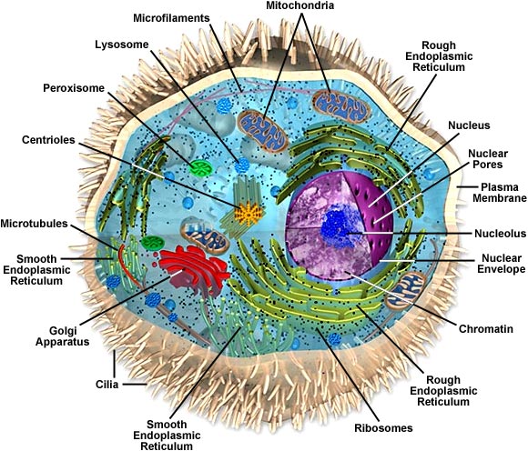
அந்தந்த தனித்தனி வேலையை செய்வதற்கு செல்லில் தனித் தனி சிறப்பு உறுப்புகள் இருக்கின்றன. அவற்றில் சில முக்கியமான உறுப்புகளை பற்றி அவைகள் என்ன செய்கின்றன என்பதை பற்றி இப்பொழுது பார்க்கலாம். (இதற்கு மேல் நீங்கள் 'செல்'லின் படத்தை பார்த்து கொண்டு மேற்கொண்டு படிப்பது உதவியாக இருக்கும்.)
பொதுவாக கண்ணுக்குத் தெரியாத மிகச் சிறிய செல் எப்படி இருக்கும் என்று கற்பனை செய்யலாம் :
அதாவது ஒரு செல்லை எடுத்துக்கொண்டு அதை ஒரு அறை அளவிற்கு பெரிதாக்கினால் பார்க்க எப்படி இருக்கும் ?

ஒரு அறை அளவிற்கு பெரிதாக மிதக்கும் ஒரு ஜெல் பந்து ஒன்றை கற்பனை செய்யுங்கள். அந்த பந்தை முழுமையாக ஒரு மெல்லிய ஊடுருவும் தன்மை கொண்ட போர்வை போத்தி இருப்பது போல கற்பனை செய்யுங்கள் அந்தப் போர்வை தான் ''செல் மெம்பரைன்". அல்லது "பிளாஸ்மா மெம்பரைன் " செல் முழுக்க பரவி இருக்கும் ஜெல் நீர் அதுதான் "சைட்டோ பிலாசம். "
அந்த ஜெல் போன்ற பந்திற்குள் உற்று பார்க்கிறீர்கள் அங்கே ஒரு கால் பந்து அளவு ஒரு அடர்த்தியான பந்து கண்ணுக்கு தெரிகிறது. அது தான் உட்கரு..நியூக்ளியஸ்.. அந்த கால் பந்துக்குள்.. x வடிவத்தில் சில பொருட்கள் மிதக்கின்றன அவைகள் தான் குரோமோசோம் பொட்டலங்கள்.( அந்த பொட்டலத்தை பிரித்து பார்த்தால் உள்ளே டி.என்.ஏ இருக்கும் , அப்புறம் அந்த DNA குள் உற்று பார்த்தால் அங்கே ஜீன்கள் இருக்கும்.)

இது தவிர அந்த கால் பந்திற்குள் இன்னும் ஒரு கிரிக்கெட் பந்து அளவு சிறிய அடர்த்தியான பந்து ஒன்று தெரிகிறது.. அதுக்கு பெயர் நியூக்ளியோலஸ். (இங்கே தான் ரிபோசோம் உற்பத்தி நடக்கிறது. அந்த ரிபோசோமின் வேலை புரோட்டீன் உண்டு பண்ணுவது.. ஒவ்வொரு செல்லுக்கும் அந்தப் புரோட்டின் இன்றியமையாதது.)
இதை தவிர அந்த ஜெல் பந்து முழுக்க பல வகை பொருட்கள் மிதக்கின்றன. மைட்டோகாண்ட்ரியா... கோல்ஜி பாடி.. லைசோசம்.. சைட்டோஸ்க்கெலட்டன்.. rough endoplasmic reticulum ,(சுருக்கமாக R.E.R என்பார்கள் ) smooth endoplasmic reticulum (S.E.R),vacuole , cytosol ,centriole. .. etc.. etc. அந்த ஒவொண்ணும் ஒவொரு வேலை...
செல்கள் பல வேலைகளை செய்கின்றன. அவற்றை சுருக்கி பார்த்தால் முக்கியமாக 3 வேலைகளை அவைகள் செய்வது தெரிய வரும். அது...
1. உணவு எடுத்து கொள்ளுதல்
2. கழிவு வெளியேற்றுதல் .
3. மறு சுழற்சி.

அப்படியே இந்த 3 செயலுக்கு உதவும் முக்கிய 3 பாகங்கள் என்ன என்று பார்த்தோமே என்றால்...
1. Plasma membrane (செல் உடலை மூடி இருக்கும் போர்வை..)
2. Nucleos (உட்கரு )
3.cytoplasm (செல் பூரா நிரம்பி உள்ள ஜெல் போன்ற திரவம் )

சரி இவற்றையும் மற்ற பாகங்களையும் ஒவொன்றாக பார்க்கலாம்...முதலில் செல் எங்கும் பரவி இருக்கும் அந்த திரவதையே எடுத்து கொள்வோம்.
Cytoplasm
இது செல் எங்கும் நிரம்பி இருக்கும் திரவம்.
சைட்டோபிளாசதின் வேலை செல் முழுக்க பொருட்களை இட பெயர்ச்சி செய்தல். அப்புறம் கார்போஹைட்ரேட் புரோட்டீன் மாதிரி பொருட்களை கரைத்து வைத்து இருத்தல்..
இது மட்டும் இல்லாமல் செல்லிற்கு உடல் வடிவத்தை இவைகள் தான் கொடுக்கின்றன. சைட்டோ பிலாசம் இல்லை எனில் செல் காற்று இறங்கிய பலூன் போல ஆகி விடும்.

Plasma_membrane :
பிளாஸ்மா மெம்பரைன் அல்லது செல் மேம்பரைன் இது முன்பே சொன்னது போல செல்லை போர்த்தி உள்ள ஒரு போர்வை ஆகும். இது எதனால் செய்ய பட்டது என்று உற்று கவனித்தீர்களேயானால்.. lipids மற்றும் proteins களால் ஆன இரட்டை அடுக்கு போர்வை இவைகள் என்பது தெரியும்.
இவைகள் எப்படி இருக்கும் என்றால்... வரிசையாக இரட்டை அடுக்கு செங்கல் அடுக்கி வைத்து ஒரு சின்ன சுவர் செய்ததை போல கற்பனை பண்ணுங்கள் அந்த இரட்டை அடுக்கு செங்கல் lipds எனும் பொருளால் ஆனது. இப்போ அந்த சின்ன சுவரில் ஒரு இடத்தில் ஒரு குட்டி கேட் ஐ கற்பனை பண்ணுங்கள் இந்த கேட் புரோட்டினால் ஆனது. கிட்டத்தட்ட இப்படி ஒரு அமைப்பினால் செய்யப்பட்ட போர்வை தான் 'செல்'லை மூடி உள்ளது. இதில் ஆக்சிஜன் மற்றும் நீர் போன்ற சிறிய மாலிகியூல்ஸ் அந்த லிபிட்ஸ் படலத்தை ஊடுருவி செல்லுக்குள் நுழைகின்றன. அதே சமயம் குளுக்கோஸ் மாதிரி பெரிய மூலக்கூறுகள் அந்த புரோட்டீன் கேட் ஐ உபயோகித்து உள்ளே நுழைகின்றன.
பிளாஸ்மா மெம்பரைன் தன்னிடம் வரும் எல்லாம் பொருட்களையும் உள்ளே அனுமதிப்பதில்லை இந்த படலம் ஒரு semi - permable படலம் அதாவது தேர்ந்தெடுக்கப்பட்ட சில பொருட்கள், மூலக்கூறுகள் , அயணிகள் தான் செல்லுக்குள் செல்ல அனுமதிக்க படுகின்றன.
அடுத்ததாக முக்கியமான ஒரு பாகத்தை பற்றி பார்க்கலாம்..
உட்கரு (nucleos ) : 
செல்லுக்குள் அடர்த்தியாக ஒரு உட்கரு காண படும் இவைகள் தான் ஒரு செல்லின் கட்டுப்பாட்டு மையம். (இவைகள் சரியாக செல்லின் மையத்தில் இருக்கும் என்று பலர் நினைப்பது உண்மை இல்லை இவைகள் செல்லில் ஓரம் சாரம் எங்கே வேணா மிதந்து கொண்டு இருக்கும் ) இந்த கட்டுப்பாட்டு மையத்தில் தான் செல்லின் மரபணு பொருட்களான DNA இருக்கும். (ஒரு செல் என்ன செய்ய வேண்டும் அதை எப்படி செய்ய வேண்டும் என்று எல்லாம் தீர்மானிப்பது இந்த டி.என்.ஏ கள்தான் )
Nucleolus 
உட்கருவிற்குள் இருக்கும் இன்னோரு பொருள் இந்த நியூகளியோலஸ். (அந்த கிரிக்கெட் பந்து..) இங்கே தான் ribosomes கள் உண்டாக்க படுகின்றன. இந்த ரிபோசோம் களின் வேலை முன்பே சொன்னது போல புரதத்தை அதாவது புரோட்டினை உண்டு பண்ணுவது. (இந்த புரோட்டினை உண்டு பண்ணுவதற்கான தகவலும் முன்பு பார்த்த DNA கிட்ட தான் இருக்கு...)
Endoplasmic_Reticulum (ER) 
ஒரு பந்தை.. நிறைய சுருக்கங்கள் நிறைந்த ஒரு துணி பையால் சுற்றியது போல.. நியூக்ளியசை சுற்றி ஒரு மடிப்பு வாய்ந்த பெட்சீட் போர்த்தியது போன்று இருக்கும் ஒரு அமைப்பு தான் எண்டோபிளாஸ்மிக் ரெடிகுலம்.. (E .R ) இந்த சுருக்கங்கள் உண்மையில் ஒரு மடிப்பு நிறைந்த பொருட்களை கடத்தும் குகை வழி. ER இன் வேலை புரோட்டினை உண்டு பண்ணும் ரைபோசோமை செல்லில் இட பெயர்ச்சி பண்ண வைப்பது. இதை rough ER என்று அழைக்கிறார்கள். காரணம் செல்லில் இன்னோரு இடத்தில் இன்னோரு ER உள்ளது அது Smooth ER இவைகள் ரிபோசோம்களை கொண்டிருப்பது இல்லை. இவைகளின் வேலை fat .. கொழுப்பை உண்டு பண்ணுவது. மற்றும் விஷ முறிவு..
Ribosomes 
மேலே சொன்ன ரைபோசம்கள் எப்படி புரதத்தை உண்டுபண்ணுகிறது என்று உற்றுப் பார்த்தால்...
அவைகள் அமினோ அமிலங்களை இணைத்து ஒரு சங்கிலித் தொடரை உருவாக்குகின்றன. இந்த அமினோ அமிலங்களின் சங்கிலித்தொடரை தான் நாம் ப்ரோடீன் அல்லது புரதம் என்கிறோம்.
(இந்த செயல்முறையை ட்ரான்ஸ்லேஷன் என்கிறார்கள். )

Golgi_body 
கோலஜி பாடியை. கோலஜி காம்ப்ளெக்ஸ் அல்லது கோலஜி அப்ரெட்ஸ் என்று வெவ்வேறு பெயர் கொண்டு அழைக்கிறார்கள். ஆனால் அதை கண்டுபிடித்தவரின் பெயரான கோல்ஜி மட்டும் மாற வில்லை. இது செல்லில் இருக்கும் இன்னோரு சுருட்டி வைக்க பட்ட பெட்சீட் அமைப்பு..
இவற்றின் வேலை என்ன தெரியுமா.. ரைபோசோம்கள் உண்டு பண்ணும் புரதம் இருக்கிறதே அது கச்சா எண்ணெய் மாதிரி பயன் படுத்த கடினமான அமைப்பில் இருக்கும் அந்த புரதத்தை வெட்டி ஒட்டி சீர் செய்து அடுக்கி வைத்து சரியாக மூட்டை கட்டி பயன் படுத்தும் வகையில் தயார் செய்து வைப்பது தான் கோல்ஜி பாடியின் வேலை. புரதத்தை சரியாக அடுக்கி வைப்பது மட்டும் இன்றி அவற்றுடன் லிபிட்ஸ் மற்றும் கார்போஹைட்ரேட்டை இணைத்து வைக்கிறது.
புரதம் கோலஜி பாடிக்கு Vesicle எனும் பார்சல் வடிவில் தான் வந்து சேர்கிறது.. அதை பயன்படுத்த கூடிய சீராக மாற்ற பட்ட புரதமாக மாற்றிய பின் அதே vesicle பார்சலில் வைத்து தான் கோலஜி அனுப்பி வைக்கிறது. அந்த vesicle அப்படியே மிதந்து போய் செல் மெம்பரைன் உடன் இணைந்து கரைந்து புரோட்டினை வெளியிடுகிறது. இதன் மூலம் எங்கே தேவையோ அந்த தேவை இருக்கும் இடத்திற்கு புரதம் கொண்டு செல்ல படுகிறது.

இப்போ அடுத்ததாக நாம் பார்க்க இருப்பது செல்லில் ஒரு மிக முக்கியமான பாகத்தை
Mitochondria 
ஒரு செல்லின் பவர் ஹவுஸ் என்று அழைக்க படுவது..இந்த மைட்டோகாண்ட்ரியா தான்.. இங்கு தான் செல்லுக்கு தேவையான ஆற்றல் உற்பத்தி நடக்கிறது. cellular respiration தான் இதன் முக்கிய வேலை. அதாவது உணவில் இருந்து பிரித்து ஆற்றலை வழங்குவது.
மைட்டோகாண்ட்ரியாவின் வேலை ATP யை உற்பத்தி செய்வது.(.. Adenosine triphosphate ) இந்த ATP என்பது செல்களுக்கு ஆற்றலை தரும் ஒரு ராசாயணம் . இது தவிர இதன் வேலை என்று பார்த்தால் செல் பிரிதல் மற்றும் செல் இறப்பில் கூட பங்கேற்கின்றன .

இவைகள் நாம் உண்ணும் உணவில் இருக்கும் வேதி ஆற்றலை ஆக்சிஜன் உதவியுடன் செல் பயன்படுத்த கூடிய ஆற்றலாக மாற்றுகின்றன. இந்த நிகழ்விற்கு பெயர் oxidative phosphorylation.
ATP என்பது ஆற்றல் வைத்து இருக்கும் பொட்டலம் எனலாம் அவைகள் தேவை படும் போது ஆற்றலை வெளியிட்டு உதவும்.

ஒரு செல் அழிவு மற்றும் புது செல் உற்பத்தி என்பது செல் சுழற்சியில் மிக முக்கியமான ஒரு தொடர்ந்து நடக்கும் செயல் .இந்த செல் அழிவிற்கு பெயர் apoptosis . ( வெவ்வேறு செல்களில் வெவ்வேறு வாழ்நாளை கொண்டிருக்கின்றன ) அதில் எந்த செல் அழிய வேண்டும் என்று தீர்மானிப்பதும் மைட்டோகாண்ட்ரியா கள் தான். மேலும் செல் அழிவில் சில என்சைம்களை கொடுத்து அழிக்க உதவுவதும் இவைகள் தான்.
ஓவல் வடிவில் இருக்கும் மைட்டோகாண்ட்ரியா பற்றி ஒரு சுவாரஸ்யமான விஷயம் சொல்கிறேன் அது ஒரு தியரி அதன் பெயர் " endosymbiyosis theory "
இதன் படி மைட்டோகாண்ட்ரியா என்பது நமது உடலுக்கு சொந்தமானதே இல்லை.. அது நமது உடலில் தங்க வந்த ஒரு பண்டைய பாக்டிரியா... கால போக்கில் அது நமது உடலின் செல்லின் ஓரு அங்கமாக மாறி விட்டது என்கிறார்கள்.
இந்த தியரிக்கு வலு சேர்க்கும் வகையில் மைட்டோகாண்ட்ரியாவின் உடலமைப்பு அமைந்து உள்ளது.

மைட்டோகாண்ட்ரியா தனக்கென தனி ரிபோசோம் கொண்டுள்ளது அதை வைத்து தனியாக புரதம் தயாரிக்கிறது. இவைகள் outer membrane ..மற்றும் innner membrane தனியாக இரண்டு அடுக்கு மெம்பரைன் கொண்டுள்ளன. அதை விட ஆச்சர்யம் மைட்டோகாண்ட்ரியா தனக்கென தனி DNA களை வைத்துள்ளது..
மேலும் மறுசுழற்சி.. மற்றும் பிரதி எடுத்தல் வேலைகளை செய்கிறது . மற்ற பாகங்கள் தன்னை பிரதி எடுக்க DNA வின் உத்தரவை கேட்க வேண்டி உள்ளது. ஆனால் மைட்டோகாண்ட்ரியா தனது சொந்த DNA தகவல் உதவி உடன் பிரதி எடுத்து கொள்கிறது. இது நியூக்ளியஸ் எனும் கட்டுப்பாடு மையத்தை நம்பி இருப்பது இல்லை. (இதனால் இதற்கு செல்ப் ரெப்ரிகேட்டிங் ஆர்கணரி என்று பெயர் ) அதாவது செல்லுக்குள் வாழும் ஒரு குட்டி செல் போல இது செயல்படுகிறது.

மைட்டோகாண்ட்ரியா இரண்டு அடுக்கு மெம்பரைன் களை கொண்டு உள்ளது அந்த இரண்டு அடுக்குகளுக்கும் நடுவே கொஞ்சம் இடைவெளி உள்ளது அந்த இடத்திற்கு பெயர் Intermembrane space
இதில் உள் மெம்பரைன் மடிப்புகள் நிறைந்ததாக உள்ளது இதை cristae என்று அழைக்கிறார்கள். அந்த உள் மடிப்பை தாண்டி நடுவில் இருக்கும் இடைவெளியில் இருப்பதை matrix என்கிறார்கள். இங்கே தான் மைட்டோகாண்ட்ரியாவின் தனி பட்ட DNA உள்ளது .மேலும் பல வகை என்சைம்கள் நிறைந்த இடம் இது. அவைகள் ATP உருவாக்கத்திற்கு முக்கிய தேவைனாவை.

மைட்டோகாண்ட்ரியா உடலில் உள்ள அனைத்து செல்களிலும் ஒரே அளவு இருக்காது.. தேவைக்கு ஏற்றார் போல இருக்கும். உதாரணமாக ஈரல் செல்கள் கொஞ்சம் அதிகம் ஆற்றல் தேவையை கொண்டு இருப்பதால் அவற்றில் மைட்டோகாண்ட்ரியாகள் 2000 திற்கும் அதிக அளவில் இருக்கும்.(. ரத்த செல்களில் இவைகள் சுத்தமாக இல்லை ) அதுவே இதய செல்களில் உள்ள சைடோபிலாசத்தில் பார்த்தால் 40 சதம் நிரம்பி இருப்பது இவைகள் தான். (அங்கு தேவை அதிகம் ) விந்தணுவில் வால் இயக்கத்திற்கு ஆற்றலை அளிக்க அங்கேயும் இவைகள் அமைந்து இருக்கின்றன.
மைட்டோகாண்ட்ரியாகள் கால்சியத்தை உறிஞ்சி வைத்து கொண்டு தேவை படும் போது பயன்படுத்துகின்றன.
மேலும் மைட்டோகாண்ட்ரியாகள் செல்லுக்கு வெப்பத்தை கூட உண்டு பண்ணி தருகின்றன.

சரி அடுத்த பாகத்திற்கு போகலாம்..
Lysosome 
நம்ம வயிற்றில் எப்படி இரைப்பை என்ற ஜீரண உறுப்பு உள்ளதோ அப்படி செல்லுக்குள் இருக்கும் ஒரு இரைப்பை என்று இதை சொல்லலாம். தனக்குள் வரும் எதையும் உடைத்து அரைத்து துண்டாக்கி விடும். உள்ளே ஒரு ஆள் அறவை இயந்திரத்துடம் காத்திருக்கும் ஒரு இடமாக லைசோசோமை கற்பனை பண்ணலாம்.
உள்ளே ஜீரணத்திற்கான சில சிறப்பு என்சைம் களை இவைகள் கொண்டு இருக்கின்றன. உனவை ஜீரணம் செய்வது ஆனாலும் சரி அல்லது இறந்த செல்களை அரைத்து துண்டாகுவதானாலும் சரி இங்கு தான் நடக்கிறது. அவ்வளவு ஏன் பாக்டிரியா மாதிரி எதிரி யாராவது ஊடுருவி விட்டால் அதையும் துண்டாகி செயல் இழக்க செய்கிறது லைசோசம்கள்.
இந்த என்சைம்களும் ரிபோசோம் இல் உண்டானவை தான். அவைகள் முன்பு பார்த்தது போல vesicle பார்சலில் கோலஜி பாடி க்கு சென்று அங்கு இறுதி வேலைகள் செய்ய பட்டு லைசோசம்களாக மாறி சைட்டோ பிலாசத்தில் மிதக்கின்றன. தேவை படும் போது செயலாற்றுகின்றன.
இவைகள் single-membrane organelles கள் ஆகும்.

எதையும் கரைக்கும் அந்த என்சைம்கள் ஏன்.. எப்படி இதன் ஒற்றை அடுக்கு மெம்பரனை கரைத்து இந்த உறுப்பையே காரைத்து ஜீரணிக்காமல் இருக்கின்றன. எப்படி இவைகள் பிழைகின்றன என்பது விஞ்ஞானிகளுக்கே ஆச்சரியமான ஒரு விஷயம். (நம்ம வயிற்றில் சுரக்கும் ஆசிட் வயிற்றை கரைப்பது இல்லை அல்லவா அப்படி தான்.. )
DNA 
உலகில் உள்ள அனைத்து வகையான மூலக்கூறுகளில் மிகவும் புகழ் வாழ்ந்த ஒரு மூலகூறு என்றால் அது deoxyribonucleic acid என்று சொல்ல கூடிய DNA தான்.
இந்த குரோமோசோம்.. என்றால் என்ன DNA என்றால் என்ன அப்புறம் ஜீன்கள் என்று எதை சொல்கிறோம் என்று பலருக்கும் குழப்பமாக இருக்கும்.. இவைகள் என்ன என்பதை பார்க்கலாம்.

செல் நியூக்ளியஸில் DNA கள் இருக்கின்றன என்று பார்த்தோம். செல்லில் உட்காருவில் உற்று பார்த்தால் அங்கே x வடிவ பொட்டலங்கள் பொட்டலம் கட்டி வைக்கப்பட்டு இருப்பதை பார்க்கலாம். இந்த x வடிவ பொட்டலங்களின் பெயர் தான் குரோமோசோம். மனித உடலில் 23 ஜோடியாக மொத்தம் 46 குரோமோசோம்கள் உள்ளன. சரி அந்தப் பொட்டலதில் என்ன இருக்கிறது என்று அதைப் பிரித்துப் பார்த்தால். உள்ளே அவைகள் குறோமேட்டின் என்கிற வலை பின்னலை வைத்து எதையோ பின்னி வைத்து இருப்பதை பார்க்கலாம். அந்த வலை பின்னலை பிரித்து உள்ளே பார்த்தால் உள்ளே அவைகள் histones என்கிற புரோட்டினால் ஆன நூலை வைத்து எதையோ சுற்றி இருப்பதை பார்க்கலாம். அந்த நூலை உரித்து எடுத்தால் உள்ளே ...அதோ சுருள் ஏணி வடிவில் பத்திரமாக இருக்கும் அந்த பொருள் தான் "DNA " . ஒவொரு குரோமோசோம்களும் தலா ஒரு DNA வை கொண்டு இருக்கும்.
ஏன் இதற்கு இத்தனை பார்சல் பாதுகாப்பு ?

உங்கள் வீட்டில் உள்ளங்கை அளவு ஒரு நூல் கண்டு இருக்கிறது என்று வைத்துக் கொள்ளுங்கள். அந்த நூலை முழுசாக பிரித்துப் போட்டுக் கொண்டே வந்தால் அந்த வீடு முழுவதும் நூல் நிரம்பிவிடும் அல்லவா. அதுவே சரியாக சுருட்டி வைக்கப்பட்டால் வீட்டில் உள்ளங்கை அளவு இடத்தை மட்டும் அது பிடித்துக்கொள்கிறது அல்லவா ? அப்படி ஒரே ஒரு செல்லில் இருக்கும் ஒரே ஒரு DNA வின் நீளம் மட்டும் கிட்ட தட்ட 3 அடி இருக்கும். மனித உடலில் மொத்தம் பல பில்லியன் கணக்கில் செல்கள் இருக்கின்றன. அவைகள் எல்லாவற்றையும் இருக்கும் டி.என்.ஏவை எடுத்து நீட்டி விட்டால் முன்பு சொன்னது போல சூரியனின் தொலைவு போல 300 மடங்கு நீளும். இதை சிறப்பாக சுருட்டி வைக்க தான் அந்த super coil அமைப்பு.
இதில் ஜீன்கள் என்பது என்ன ? அந்த DNA குள் அடங்கி இருக்கும் சில சிறப்பு தகவல் அமைப்பு தான் ஜீன்கள்.
DNA தகவல்கள் அபாரமானவை படித்து பார்த்தால்..அவற்றில் புரதம் உண்டாகுவதற்கான சமையல் குறிப்பும் உண்டு. அதே சமயம்..ஒரு மனிதனின் மொத்த தகவல் அவன் கண் என்ன நிறம் தோல் என்ன நிறம் அவன் என்ன உயரம் என்பது தொடங்கி அவனது மொத்த ஜாதகமும் அதில் உள்ளது.( அவன் ஜாதகம் மட்டுமல்ல அவன் கடந்து வந்த மொத்த பரம்பரையின் ஜாதகமும் கூட அதில் அடங்கி உள்ளது)
ஒரு குழந்தைக்கு இந்த டிஎன்ஏ தகவல் பாதி அம்மாவிடமிருந்து பாதி அப்பாவிடம் இருந்தும் கிடைக்கிறது.

DNA வில் முறுக்கிய இரண்டு இழைகளை காணலாம். அப்புறம் அவற்றிற்கு இடையில் சிறு கோடுகள் தொடர்பு கொண்டு இருப்பதை காணலாம்.
அந்த இழைகள்.. nucleotides களால் ஆனவை. அதாவது....

ஒரு phosphate மூல கூறு மற்றும்
ஒரு சர்க்கரை மூலக்கூறு. (இதன் பெயர் deoxyribose இவைகள் 5 carbons கள் அடங்கிய அமைப்பு. )
இதை தவிர ஒரு nitrogen-containing region உள்ளது அது தான் அந்த ஏணிக்கு நடுவில் உள்ள கோடுகள்.
இந்த nitrogen contenting region 4 வகைகள் கொண்டது.. அந்த 4 வகை தான் மீண்டும் மீண்டும் மாற்றி மாற்றி வரும்... அவைகள்..
adenine (A)
cytosine (C)
guanine (G) மற்றும்
thymine (T)
இந்த நான்கு எழுத்தை கொண்டு மாற்றி மாற்றி எழுத பட்ட தகவல் தான் நமது genetic code, மொத்த ஜாதகம் .
அந்த இரட்டை சூழல் ஏணி அமைப்பில் இடையில் ஒன்றோடு ஒன்று இந்த நான்கும் தொடர்பில் இருக்கின்றன. அவற்றை உற்று பார்த்தால்... A எப்போதும் T உடன் இணைவதை பார்க்கலாம்.G எப்போதும் C உடன் இணைவதை பார்க்கலாம். (அந்த நீளமான ஏணி sugar மற்றும் phosphate groups.களால் செய்ய பட்டது.)

46 குரோமோசோம் சொன்னோமே.. அது 46 கு பதில் 48 இருந்தால் என்ன ஆகும் ? அப்படி என்றால் நாம் மனிதனல்ல மங்கி. குரங்குகள் 48 க்ரோமோசோம் களை கொண்டிருக்கின்றன.
நமது குரோமோசோமில் நீண்டது முதல் குரோமோசோம் ஆகும். இதில் 8000 ஜீன்கள் உள்ளன. இருப்பதில் சிறியது 21 ஆவது குரோமோசோம் ஆகும் இதில் 3000 ஜீன்கள் உள்ளன. எப்படியும் மொத்த 46 குரோமோசோம்களில் மனிதன் மொத்தம் 20000 இல் இருந்து 30000 வரை ஜீன்களை கொண்டு இருக்கிறான்.

இந்த ஜீன்களில் தான் அந்த மனிதன் பற்றிய அனைத்து விதமான தகவல்களும் ஒளிந்திருக்கிறது. உதாரணமாக அவன் உடலில் சர்க்கரையை கட்டு படுத்தும் இன்சுலின் எனும் பொருள் சுரக்க வேண்டுமா அந்த தகவலுக்கு என்று குறிப்பிட்ட ஜீன் டேட்டா இருக்கும். இப்படி நாம் கண்டு பிடித்த தகவல் DNA வில் வெறும் 3 சதம் தான் என்கிறார்கள். மீதமுள்ள 97 சததில் ஒளிந்து இருப்பது என்னவென்று இன்னமும் நமக்கு தெரியாது
இதில் RNA என்று ஒன்றை கேள்வி பட்டு இருப்போம் அது என்ன..? இது DNA வின் காபி தான். என்ன வித்தியாசம் என்றால். இதில் இரட்டை இழைக்கு பதில் ஒற்றை இழை தான் இருக்கும். மேலும் இதில் thymine இருக்காது அதற்கு பதில் uracile என்ற ஒன்று இருக்கும். இவைகள் messenger RNA (m RNA ) ஆகும். இவைகள்
translated RNA (t RNA ) மூலம் அமினோ அமிலங்களாக டிரான்ஸ்லெட் பண்ண படுகிறது mRNA கள் three-letter sections களால் படிக்க படுகிறது அதன் பெயர் codons. ஒவொரு codon code களும்..குறிப்பிட்ட அமினோ அமிலங்களுக்கானது. (மொத்தம் கிட்ட தட்ட 20 வகை அமினோ அமிலங்கள் உள்ளன )


சரி மேலே நாம் பார்த்தது மட்டும்தான் செல்லின் பாகங்களா என்றால் இல்லை. இன்னும் Centrosome.. Peroxisome..
Villi..என்று நுணுக்கமான பல பாகங்களை செல்கள் கொண்டு இருக்கின்றன. அதில் முக்கியமான சிலதை மட்டும் தான் நாம் மேலே பார்த்தது.
மனித உடலில் உள்ள அனைத்து செல்களும் ஒரே மாதிரி இருக்காது அவைகள் தேவைக்கு தகுந்தாற்போல கொஞ்சம் மாற்றங்களுடன் காணப்படும். உதாரணமாக விந்து அணுவில் அவைகள் நீந்தி செல்ல வால் அமைப்பு இருக்கும். இந்த வாலுக்கு பெயர் "flagella ". ( இது சில பாக்டீரியாக்களிலும் இருக்கிறது.. அவைகள் நீந்தி நகர உதவுகிறது.)

இதே போல சுவாச மண்டலத்தில் எடுத்துக்கொண்டால் நுரையீரலில் முடி போன்று இருக்கும் அலை போல ஆடி கொண்டு இருக்கும் ஒரு அமைப்பு உண்டு அதன் பெயர் cillia . நாம் தூசுகளை சுவாசிக்க நேர்ந்தால். அவற்றை பிடித்து கொண்டு இரும்பும் போது தும்பும் போது வெளியேற்ற இவை உதவுகின்றன.
இப்படி உடலில் பல உறுப்புகள் பல வகைகளில் செல்களை கொண்டு இருக்கின்றன. ரத்தத்தின் செல்களும் தசையின் செல்களும் ஒரே மாதிரி இருப்பதில்லை தசைகளின் செல்களும் தோலின் செல்களும் ஒரே மாதிரி இருப்பதில்லை தோலின் செல்களும் நரம்புகளின் செல்களும் ஒரே மாதிரி இருப்பதில்லை. மொத்தம் 200 வகை இப்படி மாறுபட்ட செல்கள் மனித உடலில் இருக்கின்றன. இவைகள் அளவுகளில் கூட மாறுபட்டு இருக்கின்றன. உதாரணமாக உடலில் உள்ள மிக பெரிய அனு பெண்ணின் உடலில் உள்ள கருமுட்டையின் செல் ஆகும். என்ன ஆச்சர்யம் என்றால் இருப்பதிலேயே சிறிய செல் ஆணின் விந்து அனு செல்கள் ஆகும்.. (ஆக செல் உலக இயற்கை படி.. இருப்பதிலேயே சிறிய ஆள் ஒருவன் இருப்பதிலேயே பெரிய இடத்து ஆளை காதலித்து கரம் பிடிக்கும் செயலுக்கு பெயர் தான் குழந்தை உருவாக்கம். )
கடைசியாக சில சுவாரஸ்யமான செல் தகவல்களை சொல்லி முடிக்கிறேன்.
🥗 மனித உடலில் உள்ள செல்கள் தொடர்ந்து அழிந்து கொண்டும் புதுப்பித்துக் கொண்டும் இருக்கின்றன. ஒரு வளர்ந்த மனிதன் உடலில் ஒரே ஒரு நிமிடத்தில் கிட்ட தட்ட 9.6 கோடி செல்கள் இறந்து கொண்டு இருக்கின்றன. அதே ஒரு நிமிட நேரத்தில் 9.6 கோடி செல்கள் இரண்டாக பிரிந்து புதிய செல்கள் உண்டாகி கொண்டு இருகின்றன.
🥗 வெவ்வேறு செல்கள் வெவ்வேறு ஆயுட்காலத்தை கொண்டிருக்கின்றன. உதாரணமாக ரத்த வெள்ளை அணுக்கள் செல்கள் 13 நாள் உயிர் வாழ்கின்றன. நமது மேல் தோலில் உள்ள செல்கள் குறைந்தது ஒரு மாதம் உயிர் வாழ்கிறது. ரத்த செல்கள் குறைந்தது இரண்டு மாதங்கள் உயிர்வாழ்கின்றன. லிவரில் உள்ள செல்கள் 18 மாதங்கள் வாழ்கின்றன.
🥗 மனித உடலில் எத்தனை மனித செல்கள் உள்ளதோ அதை விடவும் அதிக எண்ணிக்கையில் பாக்டீரியா மாதிரியான மனித செல்களில் அல்லாத வேறு செல்களை மனித உடல் கொண்டிருக்கிறது.
அப்படி இருந்தும் மொத்த எடையில் வெறும் 3 சதம் தான் பாக்டிரியா மாஸ் . இது எப்படி ?? இதற்கு காரணம்..பாக்டிரியா செல்கள் (prokaryotic cell ) மனித செல்களை ஒப்பிடும் போது மிக சிறியவை.

🥗 மனிதன் பல கோடி செல்களை கொண்டு இருந்தாலும் அவனும் உருவாகும் தருணத்தில் ஒற்றை அணுவாக இருந்து தான் உண்டாகி... பல்கி பெருகி கருவாகி பல கோடி செல்கள் கொண்ட குழந்தை ஆகிறான்.
🥗 உடலில் உள்ள மிக பெரிய செல் பென்னின் கருமுட்டை என்று பார்த்தோம். வெறும் கண்ணால் பார்க்க முடிய கூடிய ஒரே செல்லும் அது தான். (ஒரு தலை முடியின் நுனி யை பார்ப்பது போல இருக்கும் ).